Subchondral bone density distribution in
Abstract | Objective: This study aims to quantitatively characterize the distribution of subchondral bone density across the human femoral head using a computed tomography derived measurement of bone density and a common reference coordinate system.
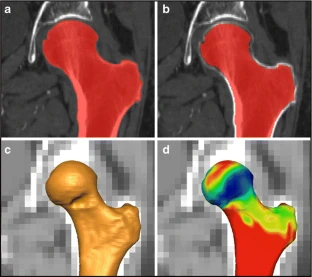
Materials and methods: Femoral head surfaces were created bilaterally for 30 patients (14 males, 16 females, mean age 67.2 years) through semi-automatic segmentation of reconstructed CT data and used to map bone density, by shrinking them into the subchondral bone and averaging the greyscale values (linearly related to bone density) within 5 mm of the articular surface. Density maps were then oriented with the center of the head at the origin, the femoral mechanical axis (FMA) aligned with the vertical, and the posterior condylar axis (PCA) aligned with the horizontal. Twelve regions were created by dividing the density maps into three concentric rings at increments of 30° from the horizontal, then splitting into four quadrants along the anterior-posterior and medial-lateral axes. Mean values for each region were compared using repeated measures ANOVA and a Bonferroni post hoc test, and side-to-side correlations were analyzed using a Pearson's correlation.
Results: The regions representing the medial side of the femoral head's superior portion were found to have significantly higher densities compared to other regions (p < 0.05). Significant side-to-side correlations were found for all regions (r(2) = 0.81 to r(2) = 0.16), with strong correlations for the highest density regions. Side-to-side differences in measured bone density were seen for two regions in the anterio-lateral portion of the femoral head (p < 0.05).
Conclusions: The high correlation found between the left and right sides indicates that this tool may be useful for understanding 'normal' density patterns in hips affected by unilateral pathologies such as avascular necrosis, fracture, developmental dysplasia of the hip, Perthes disease, and slipped capital femoral head epiphysis.

