Patient-specific finite element analysis of femurs with cemented hip implants
Abstract | Background: Over 1.6 million hip replacements are performed annually in Organisation for Economic Cooperation and Development countries, half of which involve cemented implants. Quantitative computer tomography based finite element methods may be used to assess the change in strain field in a femur following such a hip replacement, and thus determine a patient-specific optimal implant. A combined experimental-computational study on fresh frozen human femurs with different cemented implants is documented, aimed at verifying and validating the methods.
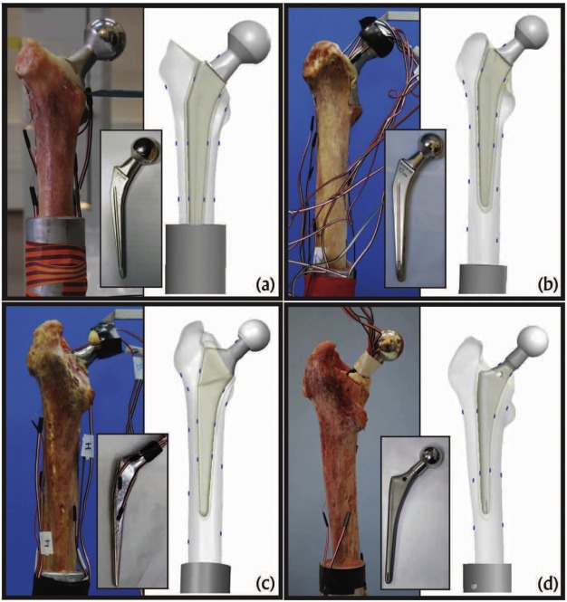
Methods: Ex-vivo experiments on four fresh-frozen human femurs were conducted. Femurs were scanned, fractured in a stance position loading, and thereafter implanted with four different prostheses. All femurs were reloaded in stance positions at three different inclination angles while recording strains on bones' and prosthesis' surfaces. High-order FE models of the intact and implanted femurs were generated based on the computer tomography scans and X-ray radiographs. The models were virtually loaded mimicking the experimental conditions and FE results were compared to experimental observations.
Findings: Strains predicted by finite element analyses in all four femurs were in excellent correlation with experimental observations FE = 1.01 × EXP – 0.07,R2 = 0.976, independent of implant's type, loading angle and fracture location.
Interpretation: Computer tomography based finite element models can reliably determine strains on femur surface and on inserted implants at the contact with the cement. This allows to investigate suitable norms to rank implants for a patient-specific femur so to minimize changes in strain patterns in the operated femur.
Keywords: Femur; P-FEMs; Total hip arthroplasty.

